


ASTM F981-04(2016)
(Practice)Standard Practice for Assessment of Compatibility of Biomaterials for Surgical Implants with Respect to Effect of Materials on Muscle and Insertion into Bone
Standard Practice for Assessment of Compatibility of Biomaterials for Surgical Implants with Respect to Effect of Materials on Muscle and Insertion into Bone
SIGNIFICANCE AND USE
4.1 This practice covers a test protocol for comparing the local tissue response evoked by biomaterials, from which medical implantable devices might ultimately be fabricated, with the local tissue response elicited by control materials currently accepted for the fabrication of surgical devices. The materials may include metals (and metal alloys), dense aluminum oxide, and polyethylene that are standardized on the basis of acceptable, well recognized, long-term response. The controls consistently produce cellular reaction and wound healing to a degree that has been found to be acceptable to the host.
SCOPE
1.1 This practice provides a series of experimental protocols for biological assays of tissue reaction to nonabsorbable biomaterials for surgical implants. It assesses the effects of the material on animal tissue in which it is implanted. The experimental protocol is not designed to provide a comprehensive assessment of the systemic toxicity, immune response, carcinogenicity, teratogenicity, or mutagenicity of the material since other standards deal with these issues. It applies only to materials with projected applications in humans where the materials will reside in bone or soft tissue in excess of 30 days and will remain unabsorbed. It is recommended that short-term assays, according to Practice F763, first be performed. Applications in other organ systems or tissues may be inappropriate and are therefore excluded. Control materials will consist of any one of the metal alloys in Specifications F67, F75, F90, F136, F138, or F562, high purity dense aluminum oxide as described in Specification F603, ultra high molecular weight polyethylene as stated in Specification F648 or USP polyethylene negative control.
1.2 This practice is a combination of Practice F361 and Practice F469. The purpose, basic procedure, and method of evaluation of each type of material are similar; therefore, they have been combined.
1.3 The values stated in SI units are to be regarded as standard. No other units of measurement are included in this standard.
1.4 This standard does not purport to address all of the safety concerns, if any, associated with its use. It is the responsibility of the user of this standard to establish appropriate safety and health practices and determine the applicability of regulatory limitations prior to use.
General Information
Buy Standard
Standards Content (Sample)
This international standard was developed in accordance with internationally recognized principles on standardization established in the Decision on Principles for the
Development of International Standards, Guides and Recommendations issued by the World Trade Organization Technical Barriers to Trade (TBT) Committee.
Designation: F981 − 04 (Reapproved 2016)
Standard Practice for
Assessment of Compatibility of Biomaterials for Surgical
Implants with Respect to Effect of Materials on Muscle and
Insertion into Bone
ThisstandardisissuedunderthefixeddesignationF981;thenumberimmediatelyfollowingthedesignationindicatestheyearoforiginal
adoption or, in the case of revision, the year of last revision.Anumber in parentheses indicates the year of last reapproval.Asuperscript
epsilon (´) indicates an editorial change since the last revision or reapproval.
1. Scope 2. Referenced Documents
2.1 ASTM Standards:
1.1 Thispracticeprovidesaseriesofexperimentalprotocols
F67 Specification for Unalloyed Titanium, for Surgical Im-
for biological assays of tissue reaction to nonabsorbable
plant Applications (UNS R50250, UNS R50400, UNS
biomaterials for surgical implants. It assesses the effects of the
R50550, UNS R50700)
material on animal tissue in which it is implanted. The
F75 Specification for Cobalt-28 Chromium-6 Molybdenum
experimental protocol is not designed to provide a comprehen-
Alloy Castings and Casting Alloy for Surgical Implants
sive assessment of the systemic toxicity, immune response,
(UNS R30075)
carcinogenicity, teratogenicity, or mutagenicity of the material
F86 Practice for Surface Preparation and Marking of Metal-
since other standards deal with these issues. It applies only to
lic Surgical Implants
materials with projected applications in humans where the
F90 Specification for Wrought Cobalt-20Chromium-
materials will reside in bone or soft tissue in excess of 30 days
15Tungsten-10NickelAlloy for Surgical ImplantApplica-
and will remain unabsorbed. It is recommended that short-term
tions (UNS R30605)
assays, according to Practice F763, first be performed. Appli-
F136 Specification for Wrought Titanium-6Aluminum-
cations in other organ systems or tissues may be inappropriate
4Vanadium ELI (Extra Low Interstitial)Alloy for Surgical
and are therefore excluded. Control materials will consist of
Implant Applications (UNS R56401)
any one of the metal alloys in Specifications F67, F75, F90,
F138 Specification for Wrought 18Chromium-14Nickel-
F136, F138,or F562, high purity dense aluminum oxide as
2.5Molybdenum Stainless Steel Bar and Wire for Surgical
described in Specification F603, ultra high molecular weight
Implants (UNS S31673)
polyethylene as stated in Specification F648 or USP polyeth-
F361 Practice for Assessment of Compatibility of Metallic
ylene negative control.
Materials for Surgical Implants with Respect to Effect of
1.2 This practice is a combination of Practice F361 and
Materials on Tissue (Withdrawn 1987)
Practice F469. The purpose, basic procedure, and method of
F469 Practice for Assessment of Compatibility of Nonpo-
evaluation of each type of material are similar; therefore, they
rous Polymeric Materials for Surgical Implants with
have been combined.
Regard to Effect of Materials on Tissue (Withdrawn
1986)
1.3 The values stated in SI units are to be regarded as
F562 Specification for Wrought 35Cobalt-35Nickel-
standard. No other units of measurement are included in this
20Chromium-10Molybdenum Alloy for Surgical Implant
standard.
Applications (UNS R30035)
1.4 This standard does not purport to address all of the
F603 Specification for High-Purity Dense Aluminum Oxide
safety concerns, if any, associated with its use. It is the
for Medical Application
responsibility of the user of this standard to establish appro-
F648 Specification for Ultra-High-Molecular-Weight Poly-
priate safety and health practices and determine the applica-
ethylene Powder and Fabricated Form for Surgical Im-
bility of regulatory limitations prior to use.
plants
1 2
ThispracticeisunderthejurisdictionofASTMCommitteeF04onMedicaland For referenced ASTM standards, visit the ASTM website, www.astm.org, or
Surgical Materials and Devices and is the direct responsibility of Subcommittee contact ASTM Customer Service at service@astm.org. For Annual Book of ASTM
F04.16 on Biocompatibility Test Methods. Standards volume information, refer to the standard’s Document Summary page on
Current edition approved April 1, 2016. Published June 2016. Originally the ASTM website.
approvedin1986.Lastpreviouseditionapprovedin2010asF981 – 04(2010).DOI: The last approved version of this historical standard is referenced on
10.1520/F0981-04R16. www.astm.org.
Copyright © ASTM International, 100 Barr Harbor Drive, PO Box C700, West Conshohocken, PA 19428-2959. United States
F981 − 04 (2016)
F763 Practice for Short-Term Screening of Implant Materi- Specifications F67, F75, F90, F138,or F562, ceramic in
als Specification F603, or polymers such as in Specification F648
polyethylene or USP Negative Control Plastic. If the test
3. Summary of Practice
materials are porous, consideration should be given to using
porous specimens for reference specimens. Alternatively, non-
3.1 This practice describes the preparation of implants, the
porous reference specimens may be used.
number of implants and test hosts, test sites, exposure
schedule, implant sterilization techniques, and methods of
6.3 Suggested Sizes and Shapes of Implants for Insertion in
implant retrieval and tissue examination of each test site.
Muscle:
Histologicalcriteriaforevaluatingtissuereactionareprovided.
6.3.1 The implants shall be cylindrical in shape and may
range from 1 mm to 6 mm in diameter and from 10 mm to 20
4. Significance and Use
mm in length depending upon the relative size of the species
4.1 This practice covers a test protocol for comparing the under study.
local tissue response evoked by biomaterials, from which 6.3.2 The dimensions used shall be reported in accordance
medical implantable devices might ultimately be fabricated,
with 8.1.
with the local tissue response elicited by control materials
6.3.3 Depending upon the particular device application,
currently accepted for the fabrication of surgical devices. The
other sample shapes may be used. For instance, an investigator
materials may include metals (and metal alloys), dense alumi-
might wish to test the biocompatibility of a new material for
num oxide, and polyethylene that are standardized on the basis
screws in the form of a screw. If an alternative specimen shape
of acceptable, well recognized, long-term response. The con-
is used, this should be reported in accordance with 8.1.
trols consistently produce cellular reaction and wound healing
6.4 Sizes and Shapes of Implants for Insertion in Bone:
to a degree that has been found to be acceptable to the host.
6.4.1 Implant diameters for use in bone shall be approxi-
mately equal to the cortex thickness. Implant lengths shall
5. Test Hosts and Sites
allow them to reside in one cortex and the medulla without
5.1 Rats (acceptable strains such as Fischer 344), New
excessive protrusion beyond the periosteum.
ZealandWhite rabbits, and other small laboratory animals may
6.4.2 The dimensions used shall be reported in accordance
be used as test hosts for soft tissue implant response. It is
with 8.1.
suggested that the rats be age and sex matched. Rabbits or
6.5 Number of Test and Control Implants:
larger animals may be used as test hosts for bone implants.
6.5.1 Ineachrat,duetosize,theremaybetwoimplants;one
Whenlargeranimalssuchasdogs,goats,orsheepareused,the
test and one control material implant.
decision should be based upon special considerations of the
6.5.2 In each rabbit, due to size, there may be six implants;
particular implant material or study.
four test and two control material implants.
5.2 The sacro-spinalis, paralumbar, gluteal muscles, and the
6.5.3 In larger animals, there may be twelve implants; eight
femur or tibia can serve as the test site for implants. However,
test material and four control material implants.
the same site must be used for test and material implants in all
6.5.4 In rabbits or larger animals, at least sixteen test
the animal species.
materials and eight materials shall be tested at each time
5.3 There shall be a minimum of four animals at each
period.
sacrifice interval for a total of twelve animals per study. If
6.6 Conditioning:
larger animals are used, in which a greater number of implants
6.6.1 Remove all surface contaminants with appropriate
may be placed, at least two animals shall be sacrificed at each
solventsandrinsealltestandcontrolimplantsindistilledwater
time period.
prior to sterilization. It is recommended that the implant
materials be processed and cleaned in the same way the final
6. Implant Specimens
product will be.
6.1 Fabrication—Each implant shall be made in a cylindri-
6.6.2 Clean, package, and sterilize all implants in the same
cal shape with hemispherical ends (see 6.3 and 6.4 for sizes).
way as used for human implantation.
If the ends are not hemispherical, this shall be reported. Each
6.6.3 Afterfinalpreparationandsterilization,handlethetest
implant shall be fabricated, finished, and its surface cleaned in
and control implants with great care to ensure that they are not
a manner appropriate for its projected application in human
scratched, damaged, or contaminated in any way prior to
subjects in accordance with Practice F86. If the specimens are
insertion.
porous, the method of preparation of the porous specimens
6.6.4 Report all details of conditioning in accordance with
shall be representative of the contemplated human implant
8.1.
application and shall yield a specimen with characteristic pore
6.7 Implantation Period—Insert all implants into each ani-
size, pore volume, and pore interconnection diameter. The
mal at the same surgical session for implantation periods of 12,
choice between using solid core specimens with porous coat-
26, and 52 weeks.
ings and specimens that are porous throughout shall be a
decision of the investigator and shall be reported.
7. Procedure
6.2 Reference metallic specimens shall be fabricated in
accordance with 6.1 from materials such as the metal alloys in 7.1 Implantation (Muscle):
F981 − 04 (2016)
7.1.1 Placematerialimplantsintheparavertebralmusclesin layer of tissue surrounding the implant. If less than a 4-mm
such a manner that they are directly in contact with muscle thick layer of tissue is removed, report in accordance with 8.1.
tissue.
7.5 Postmortem Observations—In accordance with standard
7.1.2 Introduce material implants in larger animals by the
laboratory practice, perform a necropsy on all animals that are
technique of making an implantation site in the muscle by
sacrificed for the purposes of the assay or die during the assay
using a hemostat to separate the muscle fibers. Then insert the
period. Establish the status of the health of the experimental
implant using plastic-tipped forceps or any tool that is non-
animal during the period of the assay. Report as described in
abrasive to avoid damage to the implant.
Section 8.
7.1.3 Introduce material implants using sterile technique.
7.6 Histological Procedure:
Sterile disposable needles or hypodermic tubing and trochar
7.6.1 Tissue Sample Preparation—Prepare appropriate
may be used to implant the material implants into the paraver-
blocks from each implantation site and indicate the orientation
tebral muscles along the spine. In rats, insert a negative control
of the axis of the femur relative to the axis of the implant for
implant on one side of the spine and a test material implant on
bone implants. Also indicate the orientation of the implant
the other side. In rabbits, implant one negative control material
relative to the axis of rotation of the femoral condyles.
oneachsideofthespineandimplanttwotestmaterialsoneach
7.6.1.1 Process the excised tissue block containing either a
side of the spine. If larger diameter specimens are used, an
test implant or control implant for histopathological examina-
alternative implantation technique is that described in 7.1.2.
tion and such other studies as are appropriate. Cut the sample
7.2 Implantation (Femur)—Expose the lateral cortex of
midway from end to end into appropriate size and in the
each rabbit femur and drill undersized pilot holes through the
appropriate orientation for each study. Transfer, or record, or
lateral cortex using the technique and instrument appropriate
both, the orientational details noted in 7.6.1 to each part of the
for the procedure. Final reaming of the holes should be
sample. Record the gross appearance of the implant and the
performed by hand to yield holes which are smaller than the
tissue. If the sample is porous, it is imperative that sectioning
implant specimens by 0.1 mm or less. Into each one of these
procedures be used that maintain the implant within its tissue
holes, insert one of the implants by finger pressure. Then close
envelope to allow the evaluation of tissue within the pores.
the wound.
Such procedures may include ground section preparation.
7.6.1.2 If special stains are deemed necessary, prepare
NOTE 1—Caution should be taken to minimize the motion of the
additional sections and make appropriate observations.
implant in the tissue to prevent the effects of motion on the desired result.
7.7 Histopathological Observations—Compare the amount
7.3 Postoperative Care:
of tissue reaction adjacent to the test implant to that adjacent to
7.3.1 All animal studies shall be done in a facility approved
a similar location and orientation on the control implant with
byanationallyrecognizedorganizationandinaccordancewith
respect to thickness of scar, presence of inflammatory or other
all appropriate regulations.
cell types, presence of particles, and such other indications of
7.3.2 Carefully observe each animal during the period of
interactionoftissueandmaterialasmightoccurwiththeactual
assay and report any abnormal findings.
material under test. A suggested method for the evaluation of
7.3.3 Infection or injury of the test implant site may
tissue response after implantation is Turner, et al. (1).Ifa
invalidatetheresults.Thedecisiontoreplacetheanimalsothat
porous sample is being tested, the evaluation of the tissue
the total number of retrieved implants will be as represented in
reaction shall include areas within the pores of the test and
the schedule shall be dependent upon the design of the study.
control samples at similar locations.
7.3.4 Ifananimaldiespriortotheexpecteddateofsacrifice,
7.7.1 Suggested Method for Tissue Response Evaluation:
perform a necropsy in accordance with the procedure in 7.4 to
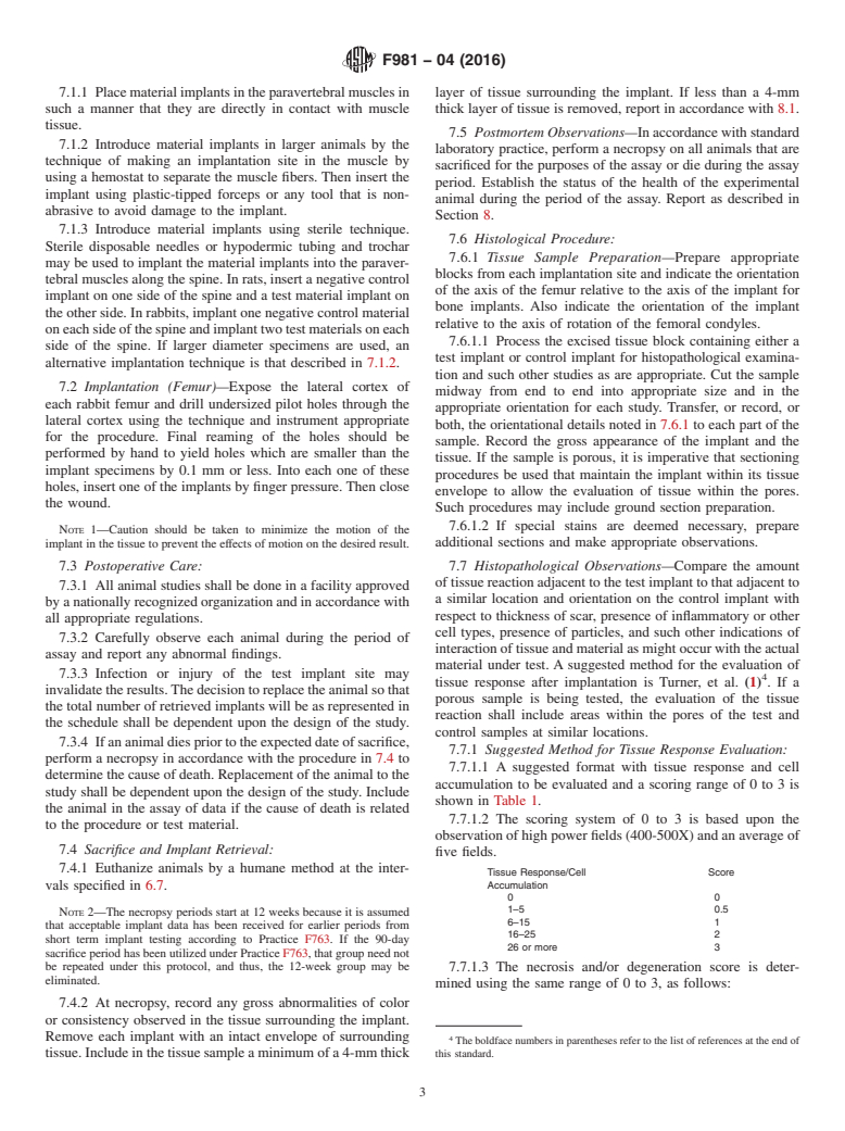
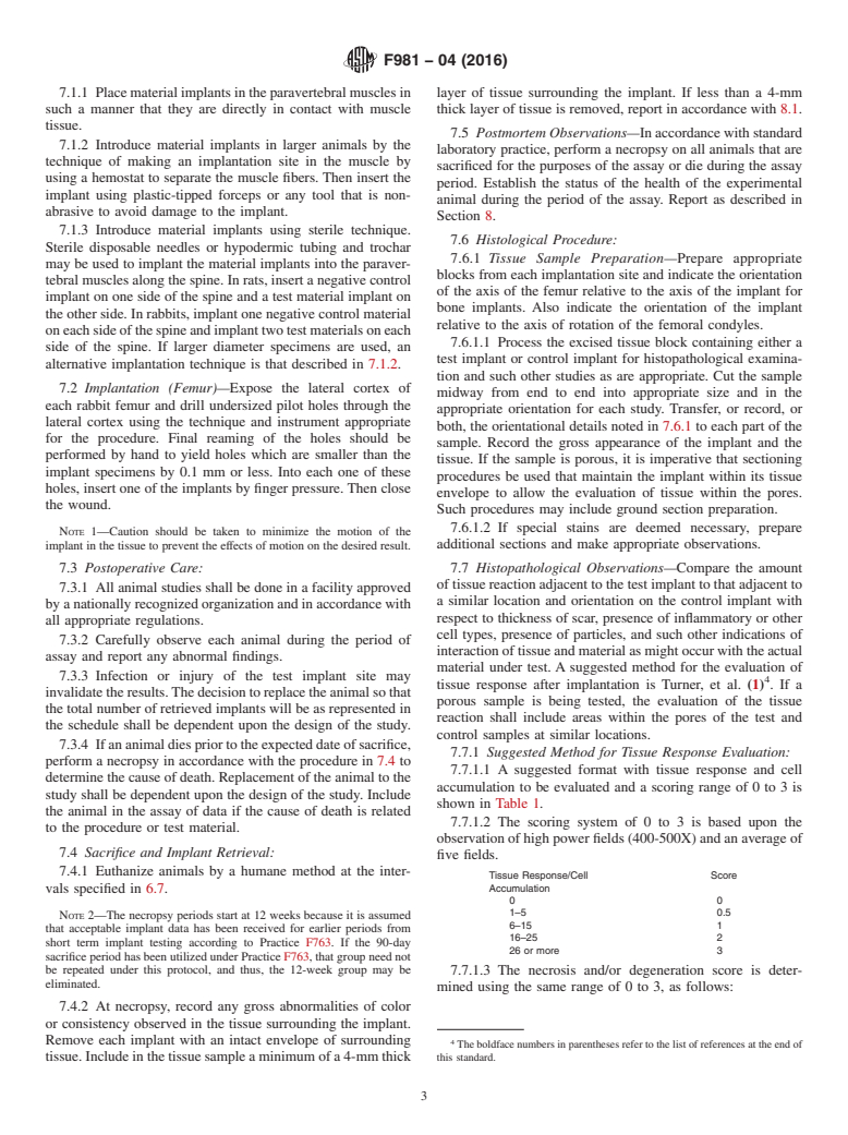
7.7.1.1 A suggested format with tissue response and cell
determine the cause of death. Replacement of the animal to the
accumulation to be evaluated and a
...
NOTICE: This standard has either been superseded and replaced by a new version or withdrawn.
Contact ASTM International (www.astm.org) for the latest information
Designation: F981 − 04 (Reapproved 2016)
Standard Practice for
Assessment of Compatibility of Biomaterials for Surgical
Implants with Respect to Effect of Materials on Muscle and
Insertion into Bone
This standard is issued under the fixed designation F981; the number immediately following the designation indicates the year of original
adoption or, in the case of revision, the year of last revision. A number in parentheses indicates the year of last reapproval. A superscript
epsilon (´) indicates an editorial change since the last revision or reapproval.
1. Scope 2. Referenced Documents
2.1 ASTM Standards:
1.1 This practice provides a series of experimental protocols
F67 Specification for Unalloyed Titanium, for Surgical Im-
for biological assays of tissue reaction to nonabsorbable
plant Applications (UNS R50250, UNS R50400, UNS
biomaterials for surgical implants. It assesses the effects of the
R50550, UNS R50700)
material on animal tissue in which it is implanted. The
F75 Specification for Cobalt-28 Chromium-6 Molybdenum
experimental protocol is not designed to provide a comprehen-
Alloy Castings and Casting Alloy for Surgical Implants
sive assessment of the systemic toxicity, immune response,
(UNS R30075)
carcinogenicity, teratogenicity, or mutagenicity of the material
F86 Practice for Surface Preparation and Marking of Metal-
since other standards deal with these issues. It applies only to
lic Surgical Implants
materials with projected applications in humans where the
F90 Specification for Wrought Cobalt-20Chromium-
materials will reside in bone or soft tissue in excess of 30 days
15Tungsten-10Nickel Alloy for Surgical Implant Applica-
and will remain unabsorbed. It is recommended that short-term
tions (UNS R30605)
assays, according to Practice F763, first be performed. Appli-
F136 Specification for Wrought Titanium-6Aluminum-
cations in other organ systems or tissues may be inappropriate
4Vanadium ELI (Extra Low Interstitial) Alloy for Surgical
and are therefore excluded. Control materials will consist of
Implant Applications (UNS R56401)
any one of the metal alloys in Specifications F67, F75, F90,
F138 Specification for Wrought 18Chromium-14Nickel-
F136, F138, or F562, high purity dense aluminum oxide as
2.5Molybdenum Stainless Steel Bar and Wire for Surgical
described in Specification F603, ultra high molecular weight
Implants (UNS S31673)
polyethylene as stated in Specification F648 or USP polyeth-
F361 Practice for Assessment of Compatibility of Metallic
ylene negative control.
Materials for Surgical Implants with Respect to Effect of
1.2 This practice is a combination of Practice F361 and
Materials on Tissue (Withdrawn 1987)
Practice F469. The purpose, basic procedure, and method of
F469 Practice for Assessment of Compatibility of Nonpo-
evaluation of each type of material are similar; therefore, they
rous Polymeric Materials for Surgical Implants with
have been combined.
Regard to Effect of Materials on Tissue (Withdrawn
1986)
1.3 The values stated in SI units are to be regarded as
F562 Specification for Wrought 35Cobalt-35Nickel-
standard. No other units of measurement are included in this
20Chromium-10Molybdenum Alloy for Surgical Implant
standard.
Applications (UNS R30035)
1.4 This standard does not purport to address all of the
F603 Specification for High-Purity Dense Aluminum Oxide
safety concerns, if any, associated with its use. It is the
for Medical Application
responsibility of the user of this standard to establish appro-
F648 Specification for Ultra-High-Molecular-Weight Poly-
priate safety and health practices and determine the applica-
ethylene Powder and Fabricated Form for Surgical Im-
bility of regulatory limitations prior to use.
plants
1 2
This practice is under the jurisdiction of ASTM Committee F04 on Medical and For referenced ASTM standards, visit the ASTM website, www.astm.org, or
Surgical Materials and Devices and is the direct responsibility of Subcommittee contact ASTM Customer Service at service@astm.org. For Annual Book of ASTM
F04.16 on Biocompatibility Test Methods. Standards volume information, refer to the standard’s Document Summary page on
Current edition approved April 1, 2016. Published June 2016. Originally the ASTM website.
approved in 1986. Last previous edition approved in 2010 as F981 – 04(2010). DOI: The last approved version of this historical standard is referenced on
10.1520/F0981-04R16. www.astm.org.
Copyright © ASTM International, 100 Barr Harbor Drive, PO Box C700, West Conshohocken, PA 19428-2959. United States
F981 − 04 (2016)
F763 Practice for Short-Term Screening of Implant Materi- Specifications F67, F75, F90, F138, or F562, ceramic in
als Specification F603, or polymers such as in Specification F648
polyethylene or USP Negative Control Plastic. If the test
3. Summary of Practice
materials are porous, consideration should be given to using
porous specimens for reference specimens. Alternatively, non-
3.1 This practice describes the preparation of implants, the
porous reference specimens may be used.
number of implants and test hosts, test sites, exposure
schedule, implant sterilization techniques, and methods of
6.3 Suggested Sizes and Shapes of Implants for Insertion in
implant retrieval and tissue examination of each test site.
Muscle:
Histological criteria for evaluating tissue reaction are provided.
6.3.1 The implants shall be cylindrical in shape and may
range from 1 mm to 6 mm in diameter and from 10 mm to 20
4. Significance and Use
mm in length depending upon the relative size of the species
4.1 This practice covers a test protocol for comparing the under study.
local tissue response evoked by biomaterials, from which
6.3.2 The dimensions used shall be reported in accordance
medical implantable devices might ultimately be fabricated, with 8.1.
with the local tissue response elicited by control materials
6.3.3 Depending upon the particular device application,
currently accepted for the fabrication of surgical devices. The
other sample shapes may be used. For instance, an investigator
materials may include metals (and metal alloys), dense alumi-
might wish to test the biocompatibility of a new material for
num oxide, and polyethylene that are standardized on the basis
screws in the form of a screw. If an alternative specimen shape
of acceptable, well recognized, long-term response. The con-
is used, this should be reported in accordance with 8.1.
trols consistently produce cellular reaction and wound healing
6.4 Sizes and Shapes of Implants for Insertion in Bone:
to a degree that has been found to be acceptable to the host.
6.4.1 Implant diameters for use in bone shall be approxi-
mately equal to the cortex thickness. Implant lengths shall
5. Test Hosts and Sites
allow them to reside in one cortex and the medulla without
5.1 Rats (acceptable strains such as Fischer 344), New
excessive protrusion beyond the periosteum.
Zealand White rabbits, and other small laboratory animals may
6.4.2 The dimensions used shall be reported in accordance
be used as test hosts for soft tissue implant response. It is
with 8.1.
suggested that the rats be age and sex matched. Rabbits or
6.5 Number of Test and Control Implants:
larger animals may be used as test hosts for bone implants.
6.5.1 In each rat, due to size, there may be two implants; one
When larger animals such as dogs, goats, or sheep are used, the
test and one control material implant.
decision should be based upon special considerations of the
6.5.2 In each rabbit, due to size, there may be six implants;
particular implant material or study.
four test and two control material implants.
5.2 The sacro-spinalis, paralumbar, gluteal muscles, and the
6.5.3 In larger animals, there may be twelve implants; eight
femur or tibia can serve as the test site for implants. However,
test material and four control material implants.
the same site must be used for test and material implants in all
6.5.4 In rabbits or larger animals, at least sixteen test
the animal species.
materials and eight materials shall be tested at each time
5.3 There shall be a minimum of four animals at each
period.
sacrifice interval for a total of twelve animals per study. If
6.6 Conditioning:
larger animals are used, in which a greater number of implants
6.6.1 Remove all surface contaminants with appropriate
may be placed, at least two animals shall be sacrificed at each
solvents and rinse all test and control implants in distilled water
time period.
prior to sterilization. It is recommended that the implant
materials be processed and cleaned in the same way the final
6. Implant Specimens
product will be.
6.1 Fabrication—Each implant shall be made in a cylindri-
6.6.2 Clean, package, and sterilize all implants in the same
cal shape with hemispherical ends (see 6.3 and 6.4 for sizes).
way as used for human implantation.
If the ends are not hemispherical, this shall be reported. Each
6.6.3 After final preparation and sterilization, handle the test
implant shall be fabricated, finished, and its surface cleaned in
and control implants with great care to ensure that they are not
a manner appropriate for its projected application in human
scratched, damaged, or contaminated in any way prior to
subjects in accordance with Practice F86. If the specimens are
insertion.
porous, the method of preparation of the porous specimens
6.6.4 Report all details of conditioning in accordance with
shall be representative of the contemplated human implant
8.1.
application and shall yield a specimen with characteristic pore
6.7 Implantation Period—Insert all implants into each ani-
size, pore volume, and pore interconnection diameter. The
mal at the same surgical session for implantation periods of 12,
choice between using solid core specimens with porous coat-
26, and 52 weeks.
ings and specimens that are porous throughout shall be a
decision of the investigator and shall be reported.
7. Procedure
6.2 Reference metallic specimens shall be fabricated in
accordance with 6.1 from materials such as the metal alloys in 7.1 Implantation (Muscle):
F981 − 04 (2016)
7.1.1 Place material implants in the paravertebral muscles in layer of tissue surrounding the implant. If less than a 4-mm
such a manner that they are directly in contact with muscle thick layer of tissue is removed, report in accordance with 8.1.
tissue.
7.5 Postmortem Observations—In accordance with standard
7.1.2 Introduce material implants in larger animals by the
laboratory practice, perform a necropsy on all animals that are
technique of making an implantation site in the muscle by
sacrificed for the purposes of the assay or die during the assay
using a hemostat to separate the muscle fibers. Then insert the
period. Establish the status of the health of the experimental
implant using plastic-tipped forceps or any tool that is non-
animal during the period of the assay. Report as described in
abrasive to avoid damage to the implant.
Section 8.
7.1.3 Introduce material implants using sterile technique.
7.6 Histological Procedure:
Sterile disposable needles or hypodermic tubing and trochar
7.6.1 Tissue Sample Preparation—Prepare appropriate
may be used to implant the material implants into the paraver-
blocks from each implantation site and indicate the orientation
tebral muscles along the spine. In rats, insert a negative control
of the axis of the femur relative to the axis of the implant for
implant on one side of the spine and a test material implant on
bone implants. Also indicate the orientation of the implant
the other side. In rabbits, implant one negative control material
relative to the axis of rotation of the femoral condyles.
on each side of the spine and implant two test materials on each
7.6.1.1 Process the excised tissue block containing either a
side of the spine. If larger diameter specimens are used, an
test implant or control implant for histopathological examina-
alternative implantation technique is that described in 7.1.2.
tion and such other studies as are appropriate. Cut the sample
7.2 Implantation (Femur)—Expose the lateral cortex of
midway from end to end into appropriate size and in the
each rabbit femur and drill undersized pilot holes through the
appropriate orientation for each study. Transfer, or record, or
lateral cortex using the technique and instrument appropriate
both, the orientational details noted in 7.6.1 to each part of the
for the procedure. Final reaming of the holes should be
sample. Record the gross appearance of the implant and the
performed by hand to yield holes which are smaller than the
tissue. If the sample is porous, it is imperative that sectioning
implant specimens by 0.1 mm or less. Into each one of these
procedures be used that maintain the implant within its tissue
holes, insert one of the implants by finger pressure. Then close
envelope to allow the evaluation of tissue within the pores.
the wound.
Such procedures may include ground section preparation.
7.6.1.2 If special stains are deemed necessary, prepare
NOTE 1—Caution should be taken to minimize the motion of the
additional sections and make appropriate observations.
implant in the tissue to prevent the effects of motion on the desired result.
7.7 Histopathological Observations—Compare the amount
7.3 Postoperative Care:
of tissue reaction adjacent to the test implant to that adjacent to
7.3.1 All animal studies shall be done in a facility approved
a similar location and orientation on the control implant with
by a nationally recognized organization and in accordance with
respect to thickness of scar, presence of inflammatory or other
all appropriate regulations.
cell types, presence of particles, and such other indications of
7.3.2 Carefully observe each animal during the period of
interaction of tissue and material as might occur with the actual
assay and report any abnormal findings.
material under test. A suggested method for the evaluation of
7.3.3 Infection or injury of the test implant site may
tissue response after implantation is Turner, et al. (1) . If a
invalidate the results. The decision to replace the animal so that
porous sample is being tested, the evaluation of the tissue
the total number of retrieved implants will be as represented in
reaction shall include areas within the pores of the test and
the schedule shall be dependent upon the design of the study.
control samples at similar locations.
7.3.4 If an animal dies prior to the expected date of sacrifice,
7.7.1 Suggested Method for Tissue Response Evaluation:
perform a necropsy in accordance with the procedure in 7.4 to
7.7.1.1 A suggested format with tissue response and cell
determine the cause of death. Replacement of the animal to the
...
This document is not an ASTM standard and is intended only to provide the user of an ASTM standard an indication of what changes have been made to the previous version. Because
it may not be technically possible to adequately depict all changes accurately, ASTM recommends that users consult prior editions as appropriate. In all cases only the current version
of the standard as published by ASTM is to be considered the official document.
Designation: F981 − 04 (Reapproved 2010) F981 − 04 (Reapproved 2016)
Standard Practice for
Assessment of Compatibility of Biomaterials for Surgical
Implants with Respect to Effect of Materials on Muscle and
Insertion into Bone
This standard is issued under the fixed designation F981; the number immediately following the designation indicates the year of original
adoption or, in the case of revision, the year of last revision. A number in parentheses indicates the year of last reapproval. A superscript
epsilon (´) indicates an editorial change since the last revision or reapproval.
1. Scope
1.1 This practice provides a series of experimental protocols for biological assays of tissue reaction to nonabsorbable
biomaterials for surgical implants. It assesses the effects of the material on animal tissue in which it is implanted. The experimental
protocol is not designed to provide a comprehensive assessment of the systemic toxicity, immune response, carcinogenicity,
teratogenicity, or mutagenicity of the material since other standards deal with these issues. It applies only to materials with
projected applications in humans where the materials will reside in bone or soft tissue in excess of 30 days and will remain
unabsorbed. It is recommended that short-term assays, according to Practice F763, first be performed. Applications in other organ
systems or tissues may be inappropriate and are therefore excluded. Control materials will consist of any one of the metal alloys
in Specifications F67, F75, F90, F136, F138, or F562, high purity dense aluminum oxide as described in Specification F603, ultra
high molecular weight polyethylene as stated in Specification F648 or USP polyethylene negative control.
1.2 This practice is a combination of Practice F361 and Practice F469. The purpose, basic procedure, and method of evaluation
of each type of material are similar; therefore, they have been combined.
1.3 The values stated in SI units are to be regarded as standard. No other units of measurement are included in this standard.
1.4 This standard does not purport to address all of the safety concerns, if any, associated with its use. It is the responsibility
of the user of this standard to establish appropriate safety and health practices and determine the applicability of regulatory
limitations prior to use.
2. Referenced Documents
2.1 ASTM Standards:
F67 Specification for Unalloyed Titanium, for Surgical Implant Applications (UNS R50250, UNS R50400, UNS R50550, UNS
R50700)
F75 Specification for Cobalt-28 Chromium-6 Molybdenum Alloy Castings and Casting Alloy for Surgical Implants (UNS
R30075)
F86 Practice for Surface Preparation and Marking of Metallic Surgical Implants
F90 Specification for Wrought Cobalt-20Chromium-15Tungsten-10Nickel Alloy for Surgical Implant Applications (UNS
R30605)
F136 Specification for Wrought Titanium-6Aluminum-4Vanadium ELI (Extra Low Interstitial) Alloy for Surgical Implant
Applications (UNS R56401)
F138 Specification for Wrought 18Chromium-14Nickel-2.5Molybdenum Stainless Steel Bar and Wire for Surgical Implants
(UNS S31673)
F361 Practice for Assessment of Compatibility of Metallic Materials for Surgical Implants with Respect to Effect of Materials
on Tissue (Withdrawn 1987)
F469 Practice for Assessment of Compatibility of Nonporous Polymeric Materials for Surgical Implants with Regard to Effect
of Materials on Tissue (Withdrawn 1986)
This practice is under the jurisdiction of ASTM Committee F04 on Medical and Surgical Materials and Devices and is the direct responsibility of Subcommittee F04.16
on Biocompatibility Test Methods.
Current edition approved June 1, 2010April 1, 2016. Published September 2010June 2016. Originally approved in 1986. Last previous edition approved in 20042010 as
F981 – 04.F981 – 04(2010). DOI: 10.1520/F0981-04R10. 10.1520/F0981-04R16.
For referenced ASTM standards, visit the ASTM website, www.astm.org, or contact ASTM Customer Service at service@astm.org. For Annual Book of ASTM Standards
volume information, refer to the standard’s Document Summary page on the ASTM website.
The last approved version of this historical standard is referenced on www.astm.org.
Copyright © ASTM International, 100 Barr Harbor Drive, PO Box C700, West Conshohocken, PA 19428-2959. United States
F981 − 04 (2016)
F562 Specification for Wrought 35Cobalt-35Nickel-20Chromium-10Molybdenum Alloy for Surgical Implant Applications
(UNS R30035)
F603 Specification for High-Purity Dense Aluminum Oxide for Medical Application
F648 Specification for Ultra-High-Molecular-Weight Polyethylene Powder and Fabricated Form for Surgical Implants
F763 Practice for Short-Term Screening of Implant Materials
3. Summary of Practice
3.1 This practice describes the preparation of implants, the number of implants and test hosts, test sites, exposure schedule,
implant sterilization techniques, and methods of implant retrieval and tissue examination of each test site. Histological criteria for
evaluating tissue reaction are provided.
4. Significance and Use
4.1 This practice covers a test protocol for comparing the local tissue response evoked by biomaterials, from which medical
implantable devices might ultimately be fabricated, with the local tissue response elicited by control materials currently accepted
for the fabrication of surgical devices. The materials may include metals (and metal alloys), dense aluminum oxide, and
polyethylene that are standardized on the basis of acceptable long-term well-characterizedacceptable, well recognized, long-term
response. The controls consistently produce cellular reaction and wound healing to a degree that has been found to be acceptable
to the host.
5. Test Hosts and Sites
5.1 Rats (acceptable strains such as Fischer 344), New Zealand White rabbits, and other small laboratory animals may be used
as test hosts for soft tissue implant response. It is suggested that the rats be age and sex matched. Rabbits or larger animals may
be used as test hosts for bone implants. When larger animals such as dogs, goats, or sheep are used, the decision should be based
upon special considerations of the particular implant material or study.
5.2 The sacro-spinalis, paralumbar, gluteal muscles, and the femur or tibia can serve as the test site for implants. However, the
same site must be used for test and material implants in all the animal species.
5.3 There shall be a minimum of four animals at each sacrifice interval for a total of twelve animals per study. If larger animals
are used, in which a greater number of implants canmay be placed, there shall be at least two animals shall be sacrificed at each
time period.
6. Implant Specimens
6.1 Fabrication—Each implant shall be made in a cylindrical shape with hemispherical ends (see 6.3 and 6.4 for sizes). If the
ends are not hemispherical, this shall be reported. Each implant shall be fabricated, finished, and its surface cleaned in a manner
appropriate for its projected application in human subjects in accordance with Practice F86. If the specimens are porous, the
method of preparation of the porous specimens shall be representative of the contemplated human implant application and shall
yield a specimen with characteristic pore size, pore volume, and pore interconnection diameter. The choice between using solid
core specimens with porous coatings and specimens that are porous throughout shall be a decision of the investigator and shall
be reported.
6.2 Reference metallic specimens shall be fabricated in accordance with 6.1 from materials such as the metal alloys in
Specifications F67, F75, F90, F138, or F562, ceramic in Specification F603, or polymers such as in Specification F648
polyethylene or USP Negative Control Plastic. If the test materials are porous, consideration should be given to using porous
specimens for reference specimens. Alternatively, nonporous reference specimens may be used.
6.3 Suggested Sizes and Shapes of Implants for Insertion in Muscle:
6.3.1 The implants shall be cylindrical in shape and may range from 1 mm to 6 mm in diameter and from 10 mm to 20 mm
in length depending upon the relative size of the species under study.
6.3.2 The dimensions used shall be reported in accordance with 8.1.
6.3.3 Depending upon the particular device application, other sample shapes may be used. For instance, an investigator might
wish to test the biocompatibility of a new material for screws in the form of a screw. If an alternative specimen shape is used, this
should be reported in accordance with 8.1.
6.4 Sizes and Shapes of Implants for Insertion in Bone:
6.4.1 Implant diameters for use in bone shall be approximately equal to the cortex thickness. Implant lengths shall allow them
to reside in one cortex and the medulla without excessive protrusion beyond the periosteum.
6.4.2 The dimensions used shall be reported in accordance with 8.1.
6.5 Number of Test and Control Implants:
6.5.1 In each rat, due to size, there may be two implants; one each test and one control material implant.
6.5.2 In each rabbit, due to size, there may be six implants; four test materials and two control material implants.
F981 − 04 (2016)
6.5.3 In larger animals, there may be twelve implants; eight test materialsmaterial and four control material implants.
6.5.4 In rabbits or larger animals, there shall be tested at least sixteen test material implants materials and eight control material
implants materials shall be tested at each time period.
6.6 Conditioning:
6.6.1 Remove all surface contaminants with appropriate solvents and rinse all test and control implants in distilled water prior
to sterilization. It is recommended that the implant materials be processed and cleaned in the same way the final product will be.
6.6.2 Clean, package, and sterilize all implants in the same way as used for human implantation.
6.6.3 After final preparation and sterilization, handle the test and control implants with great care to ensure that they are not
scratched, damaged, or contaminated in any way prior to insertion.
6.6.4 Report all details of conditioning in accordance with 8.1.
6.7 Implantation Period—Insert all implants into each animal at the same surgical session for implantation periods of 12, 26,
and 52 weeks.
7. Procedure
7.1 Implantation (Muscle):
7.1.1 Place material implants in the paravertebral muscles in such a manner that they are directly in contact with muscle tissue.
7.1.2 Introduce material implants in larger animals by the technique of making an implantation site in the muscle by using a
hemostat to separate the muscle fibers. Then insert the implant using plastic-tipped forceps or any tool that is nonabrasive to avoid
damage to the implant.
7.1.3 Introduce material implants using sterile technique. Sterile disposable needles or hypodermic tubing and trochar may be
used to implant the material implants into the paravertebral muscles along the spine. In rats, insert a negative control implant on
one side of the spine and a test material implant on the other side. In rabbits, implant one negative control material on each side
of the spine and implant two test materials on each side of the spine. If larger diameter specimens are used, an alternative
implantation technique is that described in 7.1.2.
7.2 Implantation (Femur)—Expose the lateral cortex of each rabbit femur and drill undersized pilot holes through the lateral
cortex using the technique and instrument appropriate for the procedure. Final reaming of the holes should be performed by hand
to yield holes which are smaller than the implant specimens by 0.1 mm or less. Into each one of these holes, insert one of the
implants by finger pressure. Then close the wound.
NOTE 1—Caution should be taken to minimize the motion of the implant in the tissue to prevent the effects of motion on the desired result.
7.3 Postoperative Care:
7.3.1 All animal studies mustshall be done in a facility approved by a nationally recognized organization and in accordance with
all appropriate regulations.
7.3.2 Carefully observe each animal during the period of assay and report any abnormal findings.
7.3.3 Infection or injury of the test implant site may invalidate the results. The decision to replace the animal so that the total
number of retrieved implants will be as represented in the schedule shall be dependent upon the design of the study.
7.3.4 If an animal dies prior to the expected date of sacrifice, perform a necropsy in accordance with the procedure in 7.4 to
determine the cause of death. Replacement of the animal to the study shall be dependent upon the design of the study. Include the
animal in the assay of data if the cause of death is related to the procedure or test material.
7.4 Sacrifice and Implant Retrieval:
7.4.1 Euthanize animals by a humane method at the intervals specified in 6.7.
NOTE 2—The necropsy periods start at 12 weeks because it is assumed that acceptable implant data has been received for earlier periods from short
term implant testing according to Practice F763. If the 90-day sacrifice period has been utilized under Practice F763, that group need not be repeated under
this protocol, and thus, the 12-week group may be eliminated.
7.4.2 At necropsy, record any gross abnormalities of color or consistency observed in the tissue surrounding the implant.
Remove each implant with an intact envelope of surrounding tissue. Include in the tissue sample a minimum of a 4-mm thick layer
of tissue surrounding the implant. If less than a 4-mm thick layer of tissue is removed, report in accordance with 8.1.
7.5 Postmortem Observations—In accordance with standard laboratory practice, perform a necropsy on all animals that are
sacrificed for the purposes of the assay or die during the assay period. Establish the status of the health of the experimental animal
during the period of the assay. Report as described in Section 8.
7.6 Histological Procedure:
7.6.1 Tissue Sample Preparation—Prepare appropriate blocks from each implantation site and indicate the orientation of the axis
of the femur relative to the axis of the implant (forfor bone implants).implants. Also indicate the orientation of the implant relative
to the axis of rotation of the femoral condyles.
7.6.1.1 Process the excised tissue block containing either a test implant or control implant for histopathological examination and
such other studies as are appropriate. Cut the sample midway from end to end into appropriate
...
Questions, Comments and Discussion
Ask us and Technical Secretary will try to provide an answer. You can facilitate discussion about the standard in here.